- Home
- Department
- Research
- Teaching
- Post Graduate Studies
- Services and Equipment
- Knowledge Transfer
Electron Microscopy

![]() Delibera rettorale di costituzione del CIMA
Delibera rettorale di costituzione del CIMA
The Interdepartmental Center for Advanced Microscopy Centro Interdipartimentale di Microscopia Avanzata "Carlo e Dirce Callerio" - CIMA of the University of Trieste is located in the highest part of the university district, in via Fleming 31/a and 31/b.

Electron Microscopy Service Mail
for requests to the Coordinator: cima@units.it
for technical requests / booking instruments / work sessions: cima.service@units.it
CIMA Coordinator
Dr. Gabriele Baj
e-mail: gbaj@units.it
phone +39 040 558 8676
Technical and researcher staff
Offered Services
Services are divided into the two macroareas of electron microscopy (EM) and optical microscopy (OM). Regarding EM, CIMA provides assitance and manages all stages of sample preparation, staining, and cutting. The staff has the necessary skills to determine the most appropriate techniques for sample preparation, and guide the most appropriate choice for the method of observation.
The technical staff and researchers will determine from time to time the type of digital images and/or microanalysis measurements that will be provided and possibly interpreted together with the stakeholders.
Regarding l0 OM, CIMA has adequate facilities and instrumentation to carry out classical processing/coloring/histological cytology/staining as well as to support users for live imaging acquisitions.
For users who require prolonged or repeated microscopy sessions, training is provided to enable them to use the instrumentation with varying degrees of autonomy, for observation and image acquisition.
Available instrumentation
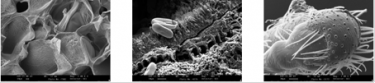
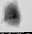
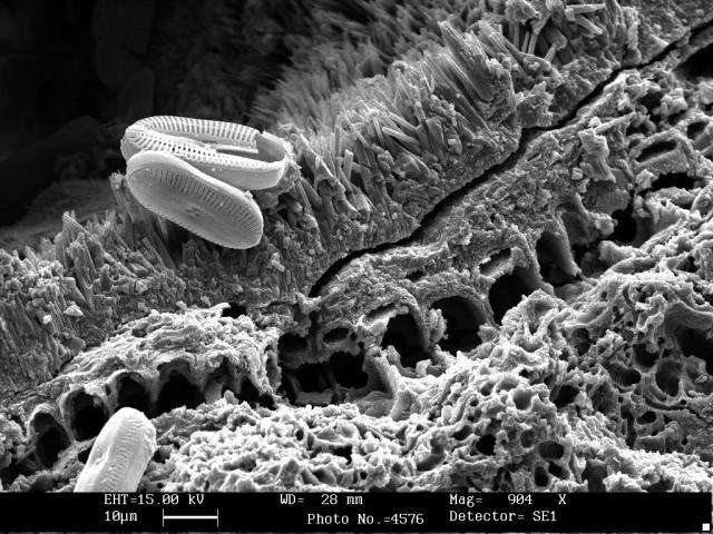
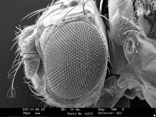
SEM
Scanning Electron Microscopy (SEM) allows high-resolution three-dimensional images to be obtained from a suitable sample. The principle on which this type of microscope is based is to send a beam of primary electrons over a conductive sample and to collect an image of the surface under investigation by converting the various resulting products into signals through appropriate detectors. These signals are then reproduced on a cathode screen whose electron brush must be in phase with that of the SEM column. The resulting image has an excellent depth of field, so that surface roughness and morphological features of the analyzed sample can be observed in detail. In addition, the analysis of the X-rays produced allows an accurate study of the elemental composition of the material under investigation.
The SEM service is equipped with the following instrumentation:
- Scanning electron microscope - GEMINI 300 FEG ZEISS
- Scanning electron microscope - Quanta 250 TI FEI
Support equipment
- S150A Sputter Coater Metallizer, whose function is to make the samples conductive
- K850 Critical Point Dryer, used when biological specimens, which are very delicate or fragile in structure, are to be dehydrated in order to observe them in vacuum
- SE imaging detector (secondary electrons)
- BSE imaging detector (backscattered electrons)
- EDX semi-quantitative microanalysis system equipped with Si(Li) PENTAFET PLUS TM detector, with ATW TM window (Oxford Instruments, United Kingdom)
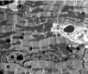
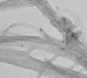
TEM
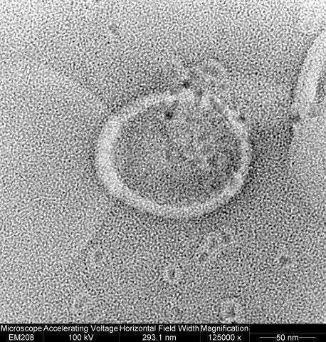
Transmission Electron Microscopy (TEM) allows obtaining , from a sufficiently thinned sample (< 0.1 microns), high-resolution images produced by high-energy electrons (up to 200 KeV) transmitted on a fluorescent screen.
The TEM service is equipped with the following instrumentation:
- Transmission electron microscope (CM 200, Philips) equipped with Quemesa camera (Olympus Soft Imaging Solutions). Image acquisition with RADIUS software.
- Transmission electron microscope (EM 208, Philips) equipped with Quemesa camera (Olympus Soft Imaging Solutions). Image acquisition with RADIUS software.
Supporting equipment.
- EDX microanalysis (Oxford Instruments, United Kingdom)
- Leica Ultramicrotome Ultracut UCT8
- Leitz Orthoplan optical microscope with bright-field, dark-field, phase-contrast condenser
- Leitz Ortholux optical microscope equipped with camera (Leica DC 300) for image acquisition

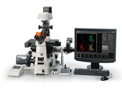
OPTICAL Microscopy (OM) allows high-magnification, high-optical-resolution imaging of specimen samples in both brightfield and fluorescence. OM also allows images to be acquired even from live (live imaging) or only minimally processed (fixed) specimens without loss of the physiological storage environment.
The OM service is equipped with the following instrumentation:
- SIM ELYRA 7 ZEISS super-resolution structured light microscope.
- A1R+ MP Nikon laser tunable multiphoton confocal microscope
- Confocal microscopy (upcoming, please refer to www.units.it/confocal for the time being)
Supporting equipment
- Dedicated cell room equipped with incubator, centrifuge, cell hood, thermostat bath
- Live imaging system for Elyra 7 microscope equipped with temperature control and CO2/O2
Access to the service & user rates
![]() Access to the service and user rates - CIMA updated to 2023
Access to the service and user rates - CIMA updated to 2023
Visits, traineeships and internships
At the facility, on request, internships / traineeships can be hosted with reference to:
- PhD students, degree students, students from graduate schools
- secondary school students who carry out internships within alternanza scuola/lavoro project
- secondary school students for visits and demonstrations
Booking calendar


Last update: 11-17-2025 - 14:26